Authors
Marco Cellani, Riccardo Pinos, Margherita Pauri, Matteo Pitton, Federica Barbaglio, AnnaSofia Tascini, Lydia Scarfò, Silvia Farè, Paolo Ghia, Cristina Scielzo.
Background
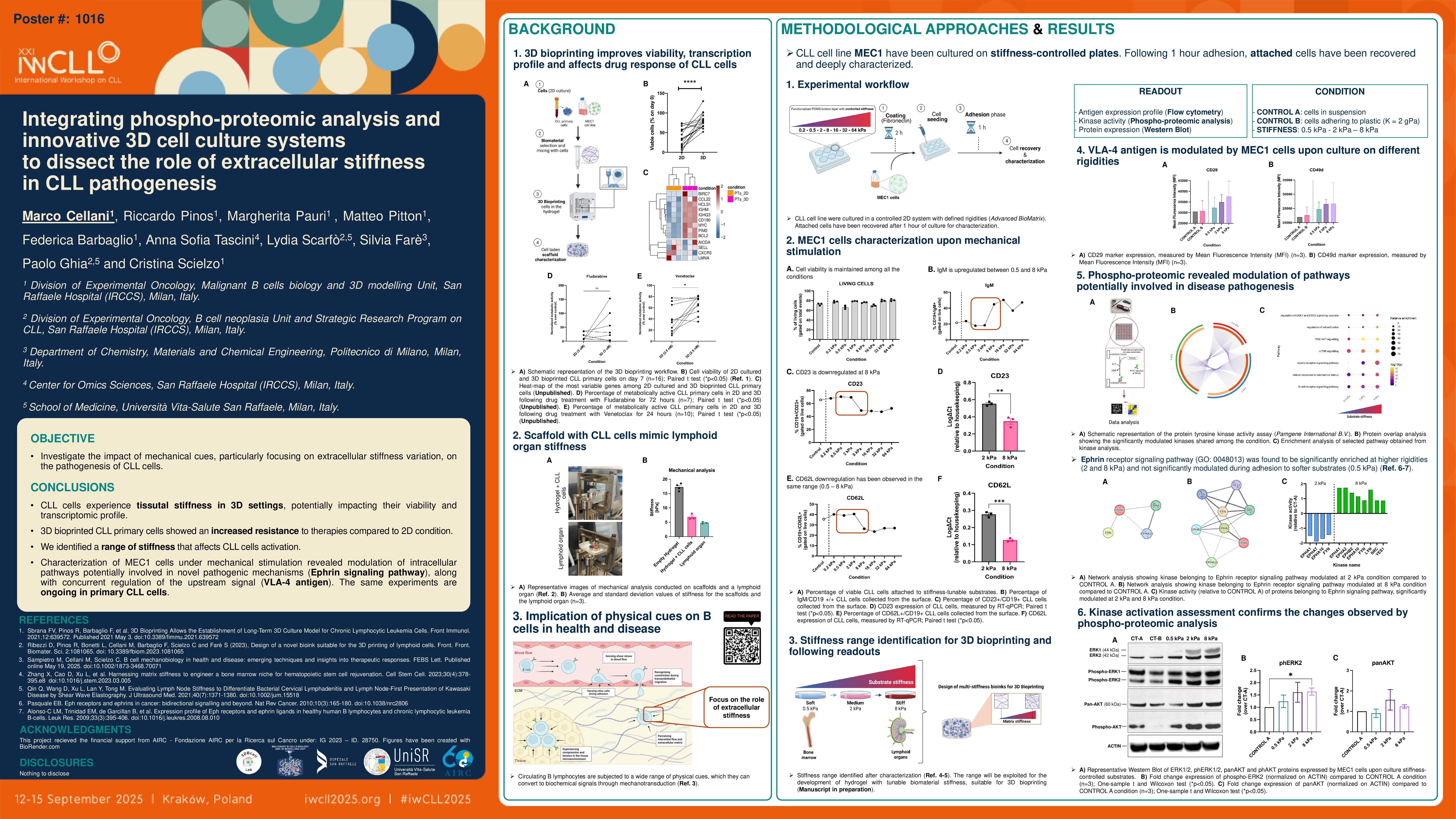
CLL is a dynamic disease recirculating between different anatomical sites in which a wide range of mechanical forces act in an orchestrated manner on malignant B lymphocytes. It is well established that cells can sense physical stimuli and convert them into biochemical signals through mechanotransduction. However, mechanobiology has been primarily explored in solid tumors, and its relevance in hematological malignancies remains poorly understood, likely due to the lack of physiologically relevant experimental models (Sampietro, Cellani et al. 2025). A deeper understanding of the implications of physical forces in the pathogenesis of CLL could pave the way for innovative strategies to control the disease. To address this gap, our research aims to investigate how mechanical stimuli influence CLL cell behavior and drug sensitivity, using both 2D and 3D culture systems, with the latter generated via 3D bioprinting. We previously demonstrated that 3D-bioprinted CLL cells can survive up to 28 days without additional stimuli, with associated changes in their gene expression profile (Sbrana et al. 2021).
Materials and methods
CLL cells were cultured in 2D on stiffness-tuned polydimethylsiloxane (PDMS)-coated plates or 3D-bioprinted in polymer-based hydrogels: CELLINK Laminink 411 (commercial) and 3A1M ink (in-house; composed of methylcellulose, alginate, and gelatin). Phenotypic responses to stiffness variation were evaluated by kinome profiling (PamGene International B.V.), western blot, and immunofluorescence. Drug response was assessed by treating both 2D-cultured and 3D-bioprinted cells with fludarabine or the BCL2 inhibitor venetoclax for 24 or 72 hours, with or without 7 days of 3D culture adaptation. Cell viability was measured by Alamar Blue assay; gene expression was analyzed by RNA-Seq and RT-qPCR.
Results
RNA-Seq performed on 3D-bioprinted CLL cells compared to 2D (n=6) revealed the modulation of markers involved in homing, adhesion, and cytoskeletal remodeling (e.g. CXCR5, PTK2, TLN1) potentially supporting CLL cells long survival. For this reason, we used this model to test the response to drugs and observed an increased resistance of 3D-bioprinted CLL cells to both chemotherapy (n=7) and targeted therapy (n=3) compared to 2D counterparts. These findings led us to reason about the mechanical characteristics of the 3D model, thus, we performed a dynamic mechanical analysis on the scaffold, showing that the latter exhibits a stiffness between 5-8 kPa, similar to the one present in lymphoid organs (Qin et al. 2020). Therefore, we hypothesized that physical cues may influence CLL cells behavior through a mechanotransduction mechanism. To test this hypothesis, we cultured CLL cells in a controlled 2D culture system using PDMS-coated plates with tunable stiffness (from 0,2 to 64 kPa). Cells proved to be viable in all the conditions and, after optimizing coating and timing of adhesion, we selected three stiffness values (0.5, 2, and 8 kPa) showing stronger variation in selected marker expression for further investigation. Following 1 hour of CLL cells adhesion to the substrate, functional kinome profiling revealed an enhanced activity of kinases involved in the ERK, PI3K/AKT and mTOR pathways at higher stiffness (8 kPa), with concurrent upregulation of the VLA-4 antigen, suggesting upstream activation triggered by a physical signal. In parallel, we observed modulation of kinases in the Ephrin family (EPHA2 and EPHB2), a group of receptor tyrosine kinases known to interact with an intricate network of pathways affecting cancer cells’ growth, migration, and invasiveness (Pasquale et al. 2010). Further experiments are ongoing to validate these pathways and to confirm their role in response to mechanical stimulation and therapy in both primary CLL cells and healthy B lymphocytes. Alongside, we are optimizing the bioprinting of hydrogels with tunable rigidities to translate findings from 2D to 3D contexts and perform drug testing in controlled stiffness settings.
Conclusions
We demonstrate that the adaptation of CLL cells to new culture settings reflects a different response to therapies and that this may be due to mechanical stimuli from the microenvironment. Functional analysis showed that stiffness influences the behaviour of CLL cells, and the pathways involved in this response are currently under investigation to confirm their prospect as mechanosensitive mechanisms, dissecting their potential involvement in CLL pathogenesis. These findings support the generation of more complex in vitro models, including the microenvironment and controlled mechanical settings, for a deeper and more reliable investigation of the mechanisms that drive CLL onset, progression, and relapse in vivo.
Keywords : Leukemia, 3D models, mechanobiology
Please indicate how this research was funded. : The project received the financial support from AIRC – Fondazione AIRC per la Ricerca sul Cancro under: IG 2023 – ID. 28750.
Please indicate the name of the funding organization.: Fondazione AIRC – Associazione Italiana per la Ricerca sul Cancro.