Authors
Bihui Pan, Jiale Zhang, Zhangdi Xu, Yu Ma, Yue Li, Jinhua Liang, Li Wang, Jiazhu Wu, Wei Xu.
Introduction
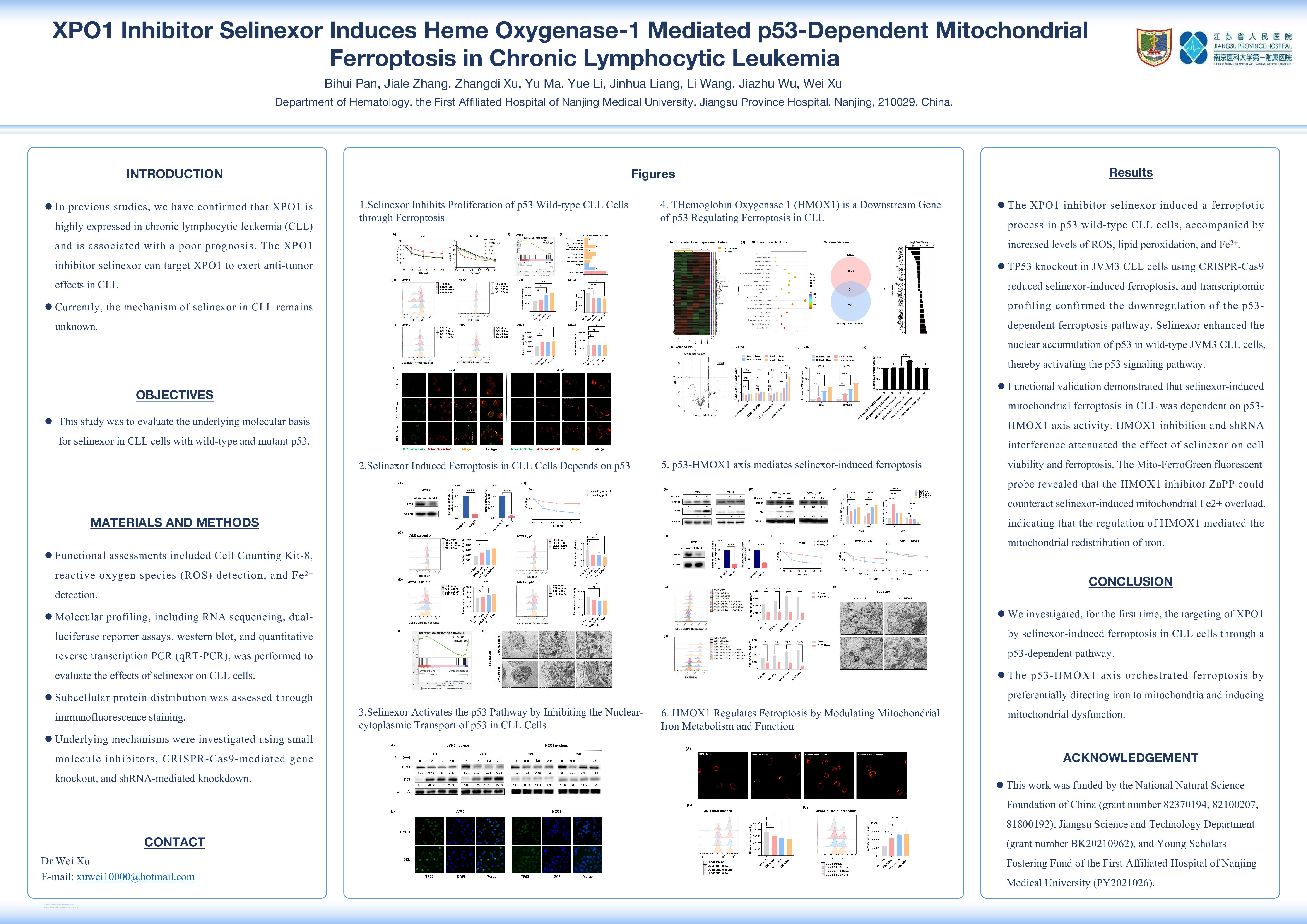
The XPO1 inhibitor selinexor can target XPO1 to exert anti-tumor effects in chronic lymphocytic leukemia (CLL). Currently, the mechanism of selinexor in CLL remains unknown.
Objectives
This study was to evaluate the underlying molecular basis for selinexor in CLL cells with wild-type and mutant p53.
Methods
Functional assessments included Cell Counting Kit-8, reactive oxygen species (ROS) detection, and Fe2+ detection. Molecular profiling, including RNA sequencing, dual-luciferase reporter assays, western blot, and quantitative reverse transcription PCR (qRT-PCR), was performed to evaluate the effects of selinexor on CLL cells. Subcellular protein distribution was assessed through immunofluorescence staining. Underlying mechanisms were investigated using small molecule inhibitors, CRISPR-Cas9-mediated gene knockout, and shRNA-mediated knockdown. In vivo, the therapeutic efficacy of selinexor was validated in NOD/SCID mouse models that monitor tumor growth.
Results
The XPO1 inhibitor selinexor induced ferroptosis in p53 wild-type CLL cells, characterized by increased ROS, lipid peroxidation, and mitochondrial Fe2+ overload, along with mitochondrial structural damage confirmed by transmission electron microscopy. This exerted anti-CLL effects both in vitro and in vivo. RNA sequencing and GSEA revealed significant enrichment of ferroptosis-related pathways in selinexor-treated JVM3 cells. TP53 knockout in JVM3 CLL cells using CRISPR-Cas9 reduced selinexor-induced ferroptosis, and transcriptomic profiling showed downregulation of the p53-dependent ferroptosis pathway. Additionally, activation of the p53 pathway by MDM2 inhibitor Nutlin-3a increased total ROS and lipid peroxidation levels in JVM3 cells. Mechanistically, selinexor enhanced nuclear p53 accumulation in wild-type JVM3 cells, activating the p53 signaling pathway and establishing p53 as a key regulator of ferroptosis in CLL. However, no reduction was observed in classic ferroptosis regulators such as SLC7A11 and GPX4 during ferroptosis in CLL cells. Through RNA-seq analysis of pre- and post-p53 knockout samples, we identified 1,539 differentially expressed genes (DEGs). Overlap with ferroptosis-related genes from the FerrDb database yielded 34 candidate downstream targets, which were individually verified based on fold change. HMOX1 was the only gene significantly upregulated under Erastin-induced ferroptosis, showing a dose-dependent effect. Furthermore, we verified the binding of the human transcription factor p53 to the HMOX1 promoter through a luciferase reporter gene assay. The results showed that p53-pcDNA3.1 significantly increased the luciferase activity of the wild-type HMOX1, demonstrating that p53 can bind to the HMOX1 promoter and increase HMOX1 transcription. Functional validation demonstrated that selinexor-induced mitochondrial ferroptosis in CLL depended on the p53-HMOX1 axis. Inhibition of HMOX1 and shRNA interference attenuated the effect of selinexor on cell viability and ferroptosis. The Mito-FerroGreen fluorescent probe revealed that ZnPP, an HMOX1 inhibitor, mitigated selinexor-induced mitochondrial Fe2+ overload, suggesting HMOX1 regulates mitochondrial iron redistribution. Immunofluorescence using specific probes for HMOX1, mitochondria, and endoplasmic reticulum demonstrated enhanced HMOX1 expression and colocalization with mitochondrial markers following selinexor treatment. Quantitative colocalization analysis confirmed substantial overlap between HMOX1 and mitochondrial signals. Furthermore, primary cells from CLL patients with different p53 mutation statuses displayed analogous responses, with selinexor inducing ferroptosis in wild-type p53 cells. In vivo, selinexor suppressed subcutaneous tumor growth in NOD/SCID mice and increased ferroptosis within tumor tissue.
Conclusion
We investigated, for the first time, the targeting of XPO1 by selinexor-induced ferroptosis in CLL cells through a p53-dependent pathway. The p53-HMOX1 axis orchestrated ferroptosis by preferentially directing iron to mitochondria and inducing mitochondrial dysfunction.
Keywords : Chronic lymphocytic leukemia; Mitochondrial ferroptosis; XPO1
Please indicate how this research was funded. : This work was funded by the National Natural Science Foundation of China (grant number 82370194) and Young Scholars Fostering Fund of the First Affiliated Hospital of Nanjing Medical University (PY2021026).
Please indicate the name of the funding organization.: National Natural Science Foundation of China and Young Scholars Fostering Fund of the First Affiliated Hospital of Nanjing Medical University