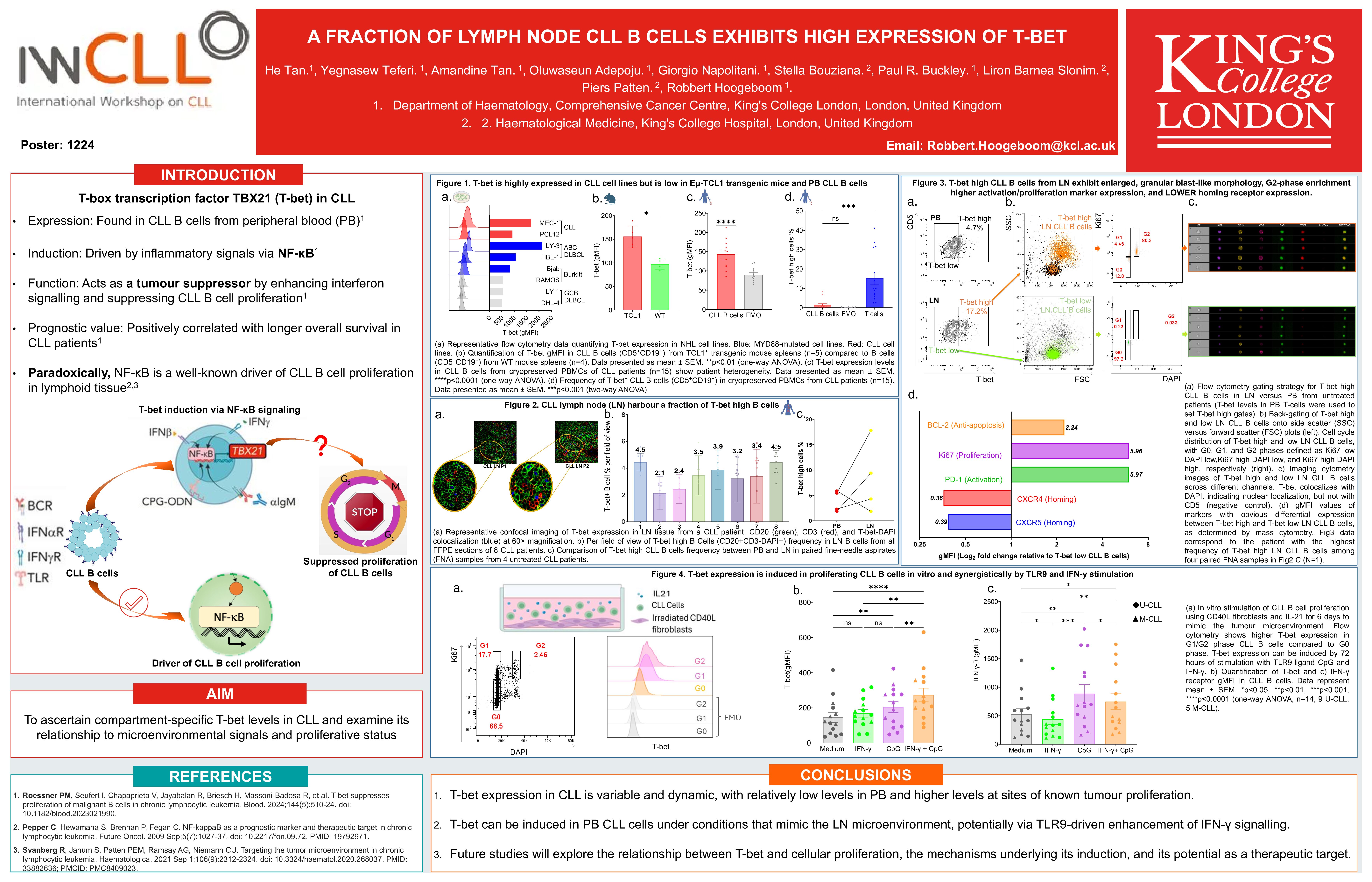
A fraction of lymph node CLL B cells exhibits high expression of T-bet (959kB pdf)
Authors
He Tan, Yegnasew Teferi, Amandine Tan, Oluwaseun Adepoju, Giorgio Napolitani, Stella Bouziana, Paul R. Buckley, Liron Barnea Slonim, Piers Patten, Robbert Hoogeboom.
Introduction
T-bet has been shown to suppress proliferation of chronic lymphocytic leukaemia (CLL) cells and expression of T-bet in peripheral blood (PB) CLL cells has been associated with increased patient survival (Roessner et al., Blood, 2024). Paradoxically, T-bet induction is driven by NF-κB activity, a known driver of CLL B cell proliferation in lymphoid tissue. We therefore set out to ascertain levels of expression of T-bet in different CLL disease compartments, and to examine how T-bet expression may relate to microenvironmental stimuli and CLL B cell proliferation.
Method
We assessed T-bet expression in CLL cell lines, a murine CLL model, and primary human CLL cells derived from PB and Lymph Nodes (LN) using qPCR, flow cytometry and multi-parameter immunofluorescence confocal microscopy, followed by investigation of the morphology and the proliferative state of T-bet high CLL B cells in primary tumour material, using conventional and imaging flow cytometry. The phenotype of T-bet high CLL subset was further characterised using a mass cytometry (CyTOF) panel containing antibodies for functional markers. Additionally, we explored mechanisms of T-bet induction by mimicking the tumour microenvironment (TME) in vitro.
Result
T-bet expression was measured in CLL and non-Hodgkin lymphoma cell lines. Elevated T-bet expression was observed in CLL cell lines (MEC-1: 3.2-fold; PCL-12: 1.8-fold), alongside activated B-cell DLBCL lines (OCI-Ly3: 4.1-fold; HBL-1: 2.0-fold), and the MYD88 L265P-mutated Burkitt’s lymphoma line Bjab (1.6-fold), normalised to germinal centre DLBCL lines (OCI-Ly1; DHL-4) and Burkitt lymphoma (Ramos) controls, where T-bet was undetectable. Quantitative PCR analysis confirmed TBX21 mRNA upregulation in CLL cell lines, consistent with T-bet protein expression trends. T-bet was also detected in PB, LN, and spleen cells derived from the Eμ-TCL1-tg mouse model for CLL.
In contrast, primary human PB CLL B cells almost exclusively exhibited intermediate T-bet expression. In cryopreserved PBMCs from CLL patients, the CD5+CD19+ CLL B cell subset displayed significantly higher T-bet protein levels relative to fluorescence minus one (FMO) control (Mean ΔgMFI= 52.72; p= 0.004), with obvious interpatient heterogeneity (p < 0.0001; n = 11). A small subset of PB CLL B cell (median frequency: 1.49%; range 0.02%–8.27%) exhibited T-bet protein levels equivalent to T-bet positive PB-derived CLL T cells (internal positive control), though their frequency remained significantly lower than T-bet high T cells (p=0.0008; n=15).
However, we identified a fraction of highly expressing T-bet CLL B cells (T-bet HI) in both formalin-fixed paraffin-embedded (FFPE) LN samples (range: 2.1–4.5%; n = 8), and LN-derived fine needle aspirates (range: 1.55–26.6%; n=4). In 3 of 4 LN samples, T-bet HI frequencies were increased compared to paired PB samples (Median fold increase: 4.7).
T-bet HI cells exhibited a larger size and enrichment in the G2 phase of the cell cycle. Mass cytometry data indicated that the T-bet HI cells co-expressed higher levels of Ki67 and BCL2, and lower levels of homing receptors (CXCR5, CXCR4). In vitro stimulation experiments demonstrated that the TLR9-ligand CpG and IFN-γ synergistically induced T-bet expression, with TLR9 signalling upregulating the IFN-γ receptor. Furthermore, induction of proliferation in vitro using CD40L+IL21 or IFN-γ+CpG gave rise to T-bet HI cells.
Conclusion
These findings indicated that levels of T-bet expression in CLL are variable and dynamic. Expression was low in blood while increased expression was observed in cells at sites of known tumour proliferation. Further, we demonstrated that T-bet can be induced in PB CLL cells under conditions that mimic the TME, potentially via TLR9-driven enhancement of IFN-γ signalling. The overall relationship between the level of T-bet expression, cellular proliferation and patient outcomes may therefore be more complex than to date explained, meriting future studies
Keywords : T-bet, CLL, Tumour-microenvironment
Please indicate how this research was funded. :
Please indicate the name of the funding organization.: