Authors
Tamar Tsertsvadze, Kurtis Edwards, Sarah John-Olabode, Irina Datikashvili-David, Ani Bilanishvili, Peter M Lydyard, Nina Porakishvili.
Introduction
We have previously shown that, differently from normal peripheral blood (PB) and tonsillar B cells, expression of the CD180 toll-like receptor is highly heterogeneous on PB and lymph node chronic lymphocytic leukaemia (CLL) cells [1-3]. The MD-1 satellite molecule, essential for CD180 expression on the cell surface, could play a role in this heterogeneity [3]. Importantly, we have shown that CD180 could hold prognostic value, since its high expression is associated with early-stage disease, M-CLL and superior overall survival [1,3]. The ability of CD180 to signal via pro-apoptotic p38MAPK pathway and re-wire IgM-mediated AKT survival signalling to a p38MAPK pathway [4], explains the positive association between CD180 expression and favourable prognosis. Similarly, higher expression of CD150, a member of the signalling lymphocyte activation molecule family (SLAMF), that is co-expressed with CD180 on CLL cells, has been associated with enhanced survival [5].
We therefore aimed to characterise the cell surface (CS) and intracellular (IC) expression of CD180 and MD-1, as well as the surface expression of CD150, in primary CLL cells and the CLL-derived MEC-1 cell line. Additionally, we assessed the correlation between these expression patterns with intracellular signalling.
Methods. Peripheral blood mononuclear cells were isolated from 26 untreated CLL patients (clinic “Aversi”, care of Dr Datikashvili-David, upon informed consent) using density gradient and immunophenotyped with APC-Cy7-conjugated anti-CD180 (clone G28-8, BD), PE-conjugated polyclonal anti-MD-1, and APC-conjugated anti-CD150 (clone SLAM.4, Abcam). CD19+ cells were identified using anti-CD19 FITC (BD). For intracellular expression, cells were stained with anti-CD19 FITC and anti-CD150 APC, fixed and permeabilized, and stained for anti-CD180 or anti-MD-1.
Eleven CD180+MD1+ CLL samples were stimulated with anti-CD180 and anti-CD150 for 30 minutes at 37°C, followed by staining with PE-conjugated anti-pAKT or APC-conjugated anti-p-p38MAPK (BD). Flow cytometry analysis was conducted on a NovoCyte 2060 (ACEA Biosciences). The same procedures were performed on the MEC1 cell line. MEC1 cells were synchronised by serum starvation and analysed at 0, 24, 48, and 72 hours.
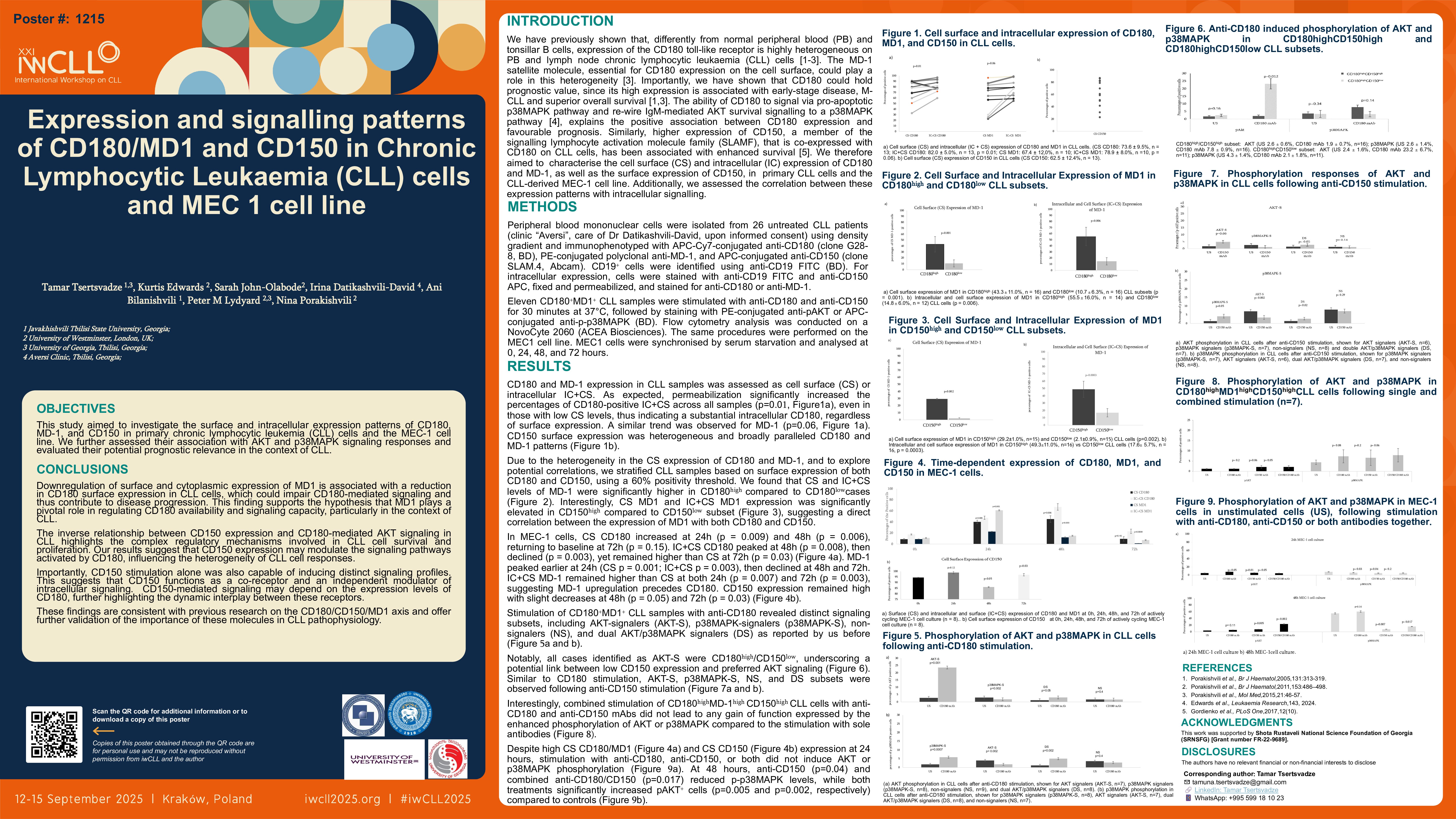
Results
CD180 and MD-1 expression in CLL samples was assessed as cell surface (CS) or intracellular (IC)+CS. As expected, permeabilization significantly increased the percentages of CD180-positive IC+CS across all samples (p=0.01), even in those with low CS levels, thus indicating a substantial intracellular CD180, regardless of surface expression. A similar trend was observed for MD-1 (p=0.06).
With the applied threshold of 60% to define positivity for csCD150 and csCD180, the levels of csMD-1-postive and cs+icMD-1-positive cells were increased in csCD180-positive (43.3±11.0% vs 10.7±6.3%; p=0.001), but also in csCD150-postive (49.3±11.0% vs 17.6±5.7%; p=0.001) cases.
Stimulation with anti-CD180 revealed distinct signalling subsets: AKT-signalers (AKT-S), p38MAPK-signalers (p38MAPK-S), non-signalers (NS), and dual AKT/p38MAPK signalers (DS). Upon anti-CD180 stimulation, CD180+CD150- CLL cells demonstrated a significant increase in p-AKT (unstimulated 2.4±1.6%, stimulated 23.2±6.7%; p=0.003), whereas p-p38MAPK remained low. In contrast, CD180+CD150+ CLL cells exhibited no AKT response, but showed slightly enhanced p38MAPK activation (unstimulated 3.6±1.4%, stimulated 7.8±1.8%, p=0.05). Like CD180, anti-CD150 stimulated CLL cells appeared to signal via preferential AKT-S, p38MAPK-S, DS or NS patterns.
In MEC-1 cells, csCD180 expression peaked at 24&48 hours before returning to baseline at 72 hours, while cs+icCD180 peaked at 48 hours and remained elevated. cs+icMD-1 peaked at 24 hours, consistently exceeding surface expression. CD150 expression remained high. MEC-1 cells did not show appreciable AKT or p38MAPK phosphorylation upon CD180 ligation. However, at 48 hours, anti-CD150 significantly reduced expression of p-p38MAPK (unstimulated 55.0±7.5%, stimulated 7.8±1.5%; p=0.002) and slightly increased p-AKT (unstimulated 3.9±0.7%, stimulated 7.5±1.7%; p=0.05). Similarly, combined CD150 and CD180 ligation resulted in reduced p-p38MAPK (unstimulated 55.0±7.5%, stimulated 10.9±4.9%; p=0.02) and increased p-AKT (unstimulated 3.9±0.9%, stimulated 23.2±19.0%; p=0.003).
Conclusion
Downregulation of MD-1 is associated with reduced CD180 surface expression, potentially weakening CD180-mediated signalling in CLL. CD150 appears to regulate CD180-induced AKT activation and also functions as an independent signalling receptor. Time-dependent expression changes observed in MEC1 cells highlight the importance of cellular context and timing in receptor-driven signalling. Overall, the CD180/MD-1/CD150 axis plays a key role in CLL pathophysiology.
1. Porakishvili et al. Br J Haematol,2005,131:313-319.
2. Porakishvili et al., Br J Haematol,2011,153:486–498.
3. Porakishvili et al., Mol Med,2015,21:46-57.
4. Edwards et al., Leukaemia Research,143, 2024.
5. Gordienko et al., PLoS One,2017,12(10).
Keywords : CD180, MD-1, CD150
Please indicate how this research was funded. : This work was supported by Shota Rustaveli National Science Foundation of Georgia (SRNSFG) [Grant number FR-22-9689].
Please indicate the name of the funding organization.: Shota Rustaveli National Science Foundation of Georgia (SRNSFG)