Authors
Šárka Pavlová, Johana Gombíková, PetrezsélyováSilvia, Macůrek Libor,Marcela Ženatová, Marie Zádrapová, Blanca Ferrer Lores, Alicia Serrano Alcalá, Eugen Tausch6, Stephan Stilgenbauer, Fanny Baran-Marszak, Gregory Lazarian, Eva Zapletalová, Jana Kopečná, Miguel Alcoceba, Ana Balanzategui, Lisa Bonello, Ivana Ježíšková, ImrichHikkel, Martina Sopkovičová, Silvia Zibellini, MarziaVarettoni, Audrey Bidet, Helena Podgornik, Sandra Šućurović, Riccardo Moia, Mónica López-Guerra, Mark Catherwood, Šárka Pospíšilová, Jitka Malčíková.
Aims
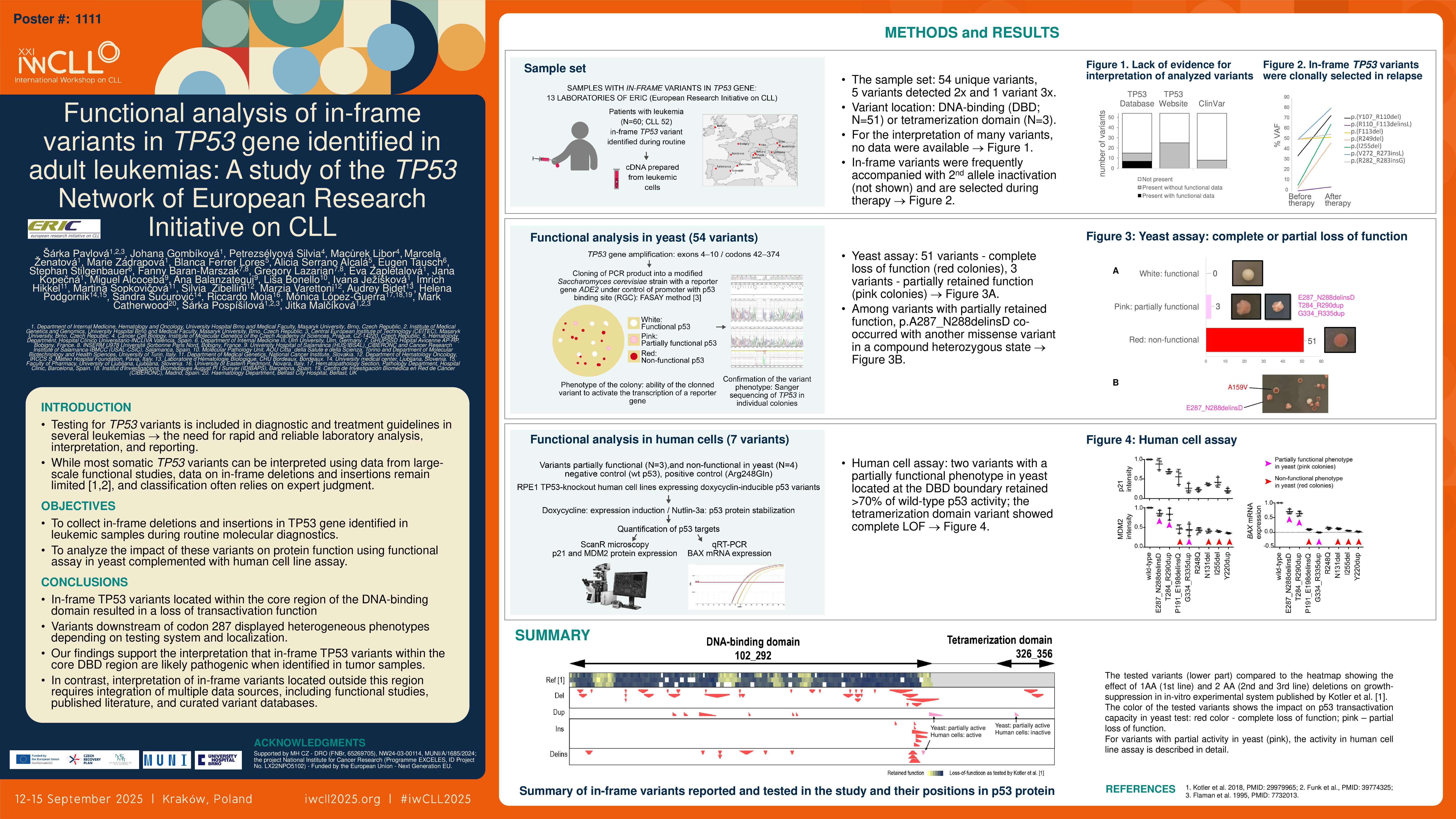
The prognostic and predictive impact of TP53 variants in leukemia has led to their inclusion in diagnostic and treatment guidelines, and increased the need for rapid and reliable laboratory analysis, interpretation, and reporting. Within its activities, European Research Initiative on CLL (ERIC) collects data on TP53 variants identified during routine assessment in CLL with the aim to create CLL-specific TP53 variant database, and addresses challenges faced by diagnostic laboratories. While most somatic TP53 variants can be interpreted using data from large-scale functional studies, data on the impact of deletions and insertions not disturbing the reading frame remain limited (Kotler et al. 2018, PMID: 29979965; Funk et al., PMID: 39774325), and classification often relies on expert judgment. Here, we approached ERIC laboratories that had identified in-frame TP53 variants and collected samples to analyze their transactivation capacity.
Methods
cDNA prepared from primary leukemic samples was used as an input material. The yeast-based Functional Analysis of Separated Alleles in Yeast (FASAY; Flaman et al. 1995, PMID: 7732013) enabled fast functional readout of individual p53 variants; variants causing loss of ability to activate ADE2 reporter gene transcription led to the accumulation of red intermediate in yeast colonies. The presence of the specific variants in yeast colonies was confirmed by Sanger sequencing. Selected variants were further tested in RPE1 TP53-knockout human cells expressing doxycyclin-inducible p53 variants, with functional assessment via nutlin-3 stabilization and measurement of endogenous MDM2 and p21 proteins and BAX mRNA levels.
Results
Sixty cDNA samples containing in-frame TP53 variants previously detected by routine gene sequencing in chronic lymphocytic leukemia (CLL; 88%) or other hematological malignancies were collected from 13 ERIC centers. The set included 54 unique variants (5 variants detected more than once). The most frequent were deletions (N=30; 1-13 amino acids), followed by delins (N=9), duplications (N=8; 1-9 amino acids), and insertions (N=7; 1-10 amino acids). The variants were mainly located in DNA-binding domain (DBD; N=51; affected codons 106 to 291) or oligomerization domain (OD; N=3; codons 336 to 350).
In FASAY, 51 variants produced red colonies, indicating a complete loss of function (LOF). Two variants located at the distal margin of the DNA-binding domain (DBD; p.Glu287_Asn288delinsAsp and p.Thr284_Arg290dup), and one in the oligomerization domain (p.Gly334_Arg335dup) yielded pink colonies, suggesting partial activity, in contrast to white colonies carrying wild-type TP53.
Testing in human cells yielded concordant results for all four selected variants that exhibited complete LOF in the yeast system. Among the variants with a partially functional phenotype in yeast, the oligomerization domain variant showed complete LOF in mammalian cells, whereas the two variants at the DBD boundary retained over 70% of wild-type p53 activity. Notably, in the original patient samples, the p.Glu287_Asn288delinsAsp variant co-occurred with a LOF missense variant in a compound heterozygous state, and the p.Thr284_Arg290dup variant was detected at variant allele frequency of only 1.9%.
Conclusion
All 49 in-frame TP53 variants located within the core region of the DNA-binding domain resulted in a loss of transactivation function, while the variants downstream of codon 287 displayed heterogeneous phenotypes depending on testing system and localization, with variants located in OD being predominantly non-functional. Our findings support the interpretation that in-frame TP53 variants within the core DBD region are likely pathogenic when identified in tumor samples. In contrast, interpretation of in-frame variants located outside this region requires integration of multiple data sources, including functional studies, published literature, and curated variant databases.
Supported by MH CZ – DRO (FNBr, 65269705), NW24-03-00114, MUNI/A/1685/2024; the project National Institute for Cancer Research (MEYS, Programme EXCELES, ID Project No. LX22NPO5102) – co-funded by the European Union – Next Generation EU.
Keywords : in-frame, TP53, mutation
Please indicate how this research was funded. : Supported by MH CZ – DRO (FNBr, 65269705), NW24-03-00114, MUNI/A/1685/2024; the project National Institute for Cancer Research (MEYS, Programme EXCELES, ID Project No. LX22NPO5102) – co-funded by the European Union – Next Generation EU.
Please indicate the name of the funding organization.: Ministry of health of the Czech republic, Czech health research council, The Ministry of Education, Youth and Sports (MSMT) and European Union