Authors
Martina Cardillo, PhD, Matthew J Friedman,BS, Anita Ng, PhD, Randall S. Davis, MD, Xiao J Yan, Ashna Shah, BS, Michael Ryan, Peter K Gregersen, MD, Steven L. Allen,MD, Jonathan E Kolitz, MD, Kanti R. Rai,MD, Paolo Ghia,MD PhD, Nicholas Chiorazzi,MD.
T-cell activation via T-cell receptor (TCR) signaling is less robust in patients with chronic lymphocytic leukemia (CLL) than in healthy controls (HC), contributing to the immune deficiency characteristic of CLL. In HCs, activated B cells are potent antigen-presenting cells (APCs), initiating T-cell responses through the presentation of processed antigenic peptides via MHC class II. However, in CLL, this process, particularly immune synapse formation, is defective. Most CLL cells and unswitched normal B cells express surface membrane IgM (smIgM) and IgD (smIgD), which share identical antigen-binding sites, and each isotype participates in antigen uptake. However, whether they do so equivalently and activate T cells equally have not been directly tested. Therefore, we evaluated if selectively targeting a foreign antigen to smIgM or smIgD affected antigen internalization, processing, and T-cell activation in HCs and people with CLL.
We engineered two murine monoclonal antibodies (mAbs) that are specific for human IgM or IgD and have very similar affinity constants for the respective antigens to express human IgG4 Fc domains (to avoid Fc receptor binding) and either included or excluded the receptor-binding domain (RBD) of the SARS-CoV-2 spike protein to the Fc domain: r-αIgM, r-αIgM-RBD, r-αIgD, and r-αIgD-RBD. The CXCR4DimCD5Bright CLL subset, also known as the proliferative fraction (PF), which is enriched for antigen presentation molecules, was the target of antigen delivery, uptake, and presentation. T-cell activation was measured by flow cytometry based on the enhanced expression of CD69, CD134, CD137, and the presence of intracellular IFNγ and IL-4. Confocal microscopy using the r-αIgM and r-αIgD conjugated with AF488 or AF647 or with pHrodo-labeled mAbs (Deep Red for IgM, Green for IgD) was used to track internalization and compartment deposition.
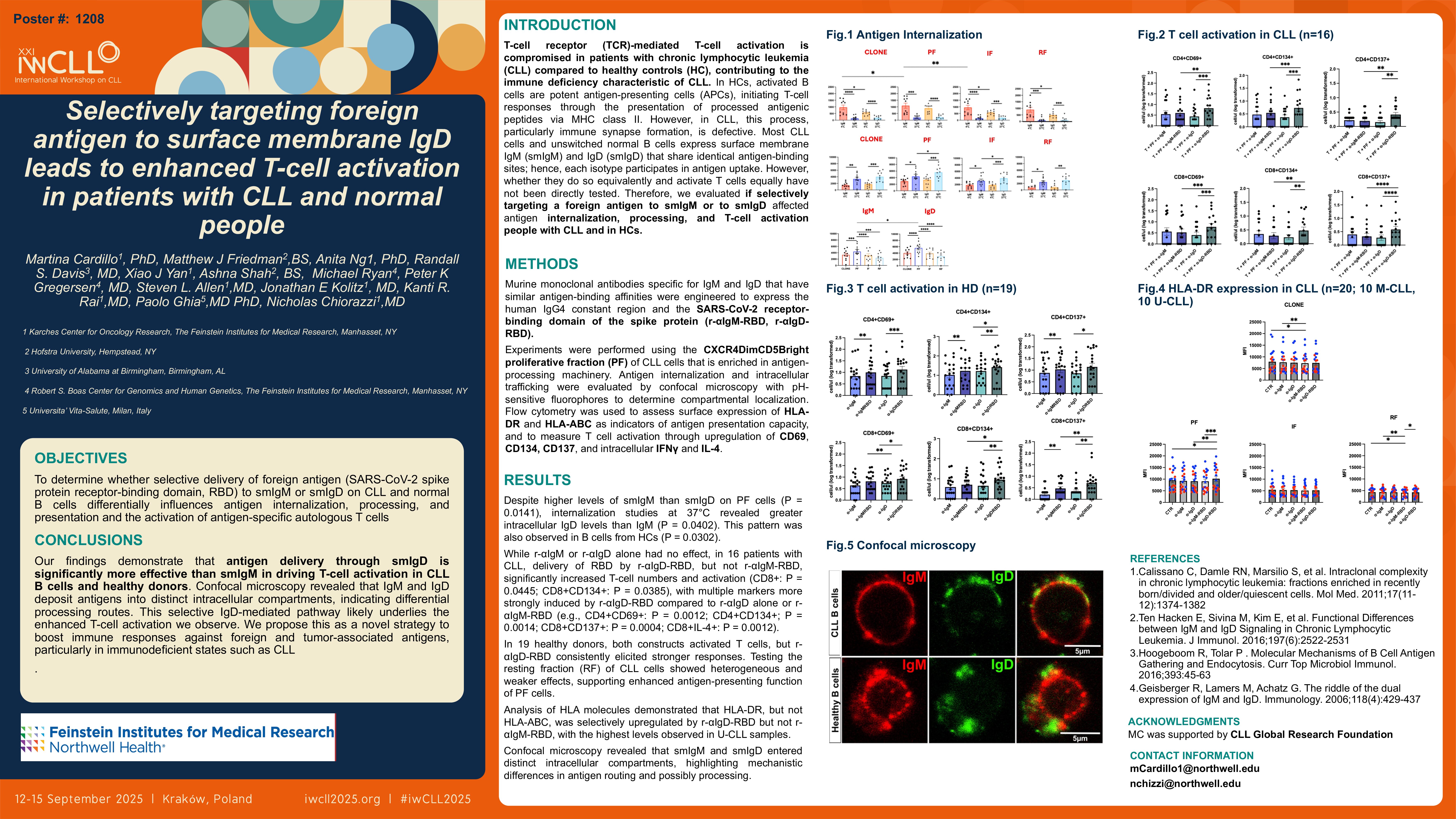
Despite higher smIgM than smIgD expression on PF cells (P = 0.0141), internalization at 37°C showed significantly greater intracellular IgD than IgM (P = 0.0402), a pattern also observed in normal B cells (P = 0.0302).
While treatment with r-αIgM or r-αIgD alone had no impact, delivery of RBD via r-αIgD-RBD, but not via r-αIgM-RBD, resulted in significantly enhanced T-cell proliferation and activation (CD8+ P = 0.0445; CD8+CD134+ P = 0.0385). Numerous activation markers were significantly upregulated with r-αIgD-RBD versus r-αIgD alone, and also when directly compared to r-αIgM-RBD (e.g., CD4+CD69+ P=0.0012; CD4+CD134+ P=0.0014; CD8+CD137+ P = 0.0004; CD8+IL-4+ P = 0.0012; ). To validate these findings in a physiologic setting, we tested B cells from 19 healthy donors using the same antibody panel. In contrast to the CLL setting, both r-αIgM-RBD and r-αIgD-RBD stimulated T-cell responses, although r-αIgD-RBD consistently elicited stronger T-cell activation than r-αIgM-RBD across multiple readouts. Hence, even for normal B cells, smIgD mediates a more immunostimulatory outcome.
To ensure that this effect was specific to the PF and not generalizable across all CLL cells, we tested the resting fraction (RF, CXCR4BrightCD5Dim) in 10 patients. In contrast to the consistent PF data, the RF results were heterogeneous. While some patients showed responsiveness to r-αIgM-RBD, overall T-cell activation was less consistent and not as robust as with the PF. These findings are consistent with antigen-presenting capacity varying with the activation state of B cells and PF cells being more effective for modeling immunostimulatory mechanisms.
To further explore the mechanistic basis for these differential responses, we evaluated HLA-DR expression, an essential component of antigen presentation, in 20 CLL samples (10 mutated [M-CLL] and 10 unmutated [U-CLL]). After 1-hour incubation with the r-αIgM-RBD or r-αIgD-RBD, HLA-DR expression was significantly higher in the PF compared to the RF and was significantly enhanced only by r-αIgD-RBD. Notably, the highest levels of surface HLA-DR expression were observed in U-CLL samples, suggesting that these cells may have a greater intrinsic capacity for antigen presentation.
Finally, we used confocal microscopy to identify the intracellular locations of IgM and IgD after surface membrane engagement. Remarkably, this revealed that smIgM and smIgD arrive in distinct intracellular compartments.
Collectively, these findings suggest that smIgD elicits more effective antigen processing and presentation, resulting in greater activation of foreign antigen-specific T cells in both CLL and healthy individuals. This distinct smIgD-mediated pathway may offer a novel strategy to enhance immune responses to foreign and tumor-associated antigens, particularly in immunodeficient states like CLL.
Keywords : leukemia, BCR, IgD
Please indicate how this research was funded. :
Please indicate the name of the funding organization.: CLL Global Research Foundation